- Experimental Study of MEBT/MEBO on the Pathological Changes of Rat Refractory Wound Model

-
目录
- 掌握再生生命定律 争做一代医学大师
- Master the Law of Regenerated Life and Try to Be a Medical Grandmaster of Time
- MEBT/MEBO对大鼠慢性难愈合模型创面病理学改变影响的实验研究
- Experimental Study of MEBT/MEBO on the Pathological Changes of Rat Refractory Wound Model
- MEBO与SD-Ag乳膏治疗放射性烧伤临床对比研究
- 烧伤病房耐甲氧西林金黄色葡萄球菌mec耐药基因及HVR基因分型研究
- 湿润烧伤膏的临床新用
- 湿润半暴露疗法治疗耳廓深Ⅱ度烧伤临床观察
- MEBO治疗成人阴囊阴茎深度烧伤
- 湿性薄化技术治疗深度烧伤35例疗效观察
- 预防HIV阳性烧伤患者职业暴露的体会
- 烧伤患者心理状况调查及健康指导
- 注射壶外置皮肤软组织扩张术的临床护理
- 美宝创疡贴治疗皮肤挫擦伤35例临床观察
- 应用90Se敷贴治疗瘢痕疙瘩术后复发的临床观察
- 湿性医疗技术配合中药药浴治疗糖尿病足湿性坏疽
- 美宝创疡贴治疗下肢慢性溃疡16例
- 应用美宝创疡贴加湿润烧伤膏治疗难愈性溃疡的临床总结
- 美宝创疡贴在褥疮护理应用中的体会
- 美宝湿润烧伤膏治疗鼻黏膜糜烂的体会
- MEBO治疗大疱性表皮松解型药疹临床体会
- 白介素-21趋化因子受体CCR9辅助T细胞亚群选择性影响消化系统的附属器官
- CCR9+ Th cells selectively affect accessory organs of the digestive system
TANG Qian-li, FU Jun, DI Jia-qi, ZHAO Shu-xiao, HAN Shan-shan, DAI Bo, FENG JingFundation: National Natural Science Fundation Project of 2008 (No.: 30860356); Scientific study subject of Guangxi University of TUN of 2009 (No.: P2009132)
Affiliations: Guangxi University of TCM, Nanning 530001, Guangxi Province, China(TANG Qian-li, FU Jun, DI Jia-qi,HAN Shan-shan, DAI Bo, FENG Jing ); The First Affiliated Hospital of Guangxi University of TCM, Nanning 530023, Guangxi Province, China(ZHAO Shu-xiao).
【Abstract】 Objective To explore the influence and the probable mechanism of MEBT/MEBO on the granulation tissue of rat model with refractory wound. Methods: A total of 45 SPF grade SD male rats were randomly divided into three groups: MEBO group, Recombinant Bovine Basophilic Fibroblast Growth Factor (RBBFGF) group, and model group, with 15 rats in each group. The healing status of wounds was observed on the 8th day of the interventions and the following were analyzed: i. Growth of granulation tissue; ii. tissue condition of wound under the optimal microscope; iii. tissue condition of wound under the electron microscope. Results: The healing time of MEBO group was 20.13±1.41days, less than that of RBBFGF group (22.07±1.62 days), and apparently less than that of model group (30.40±1.18 days). All results had statistical significance (P<0.01); The examination of optical microscope indicated: Number of capillaries in MEBO group was more than that of RBBFGF group and model group. The numbers of capillary are 12.13±2.26, 9.87±1.99, 7.60±1.50 respectively in MEBO group, RBBFGF group and model group. When MEBO group was compared to RBBFGF group and model group, there was a statistical significance(P<0.01). The examination of electron microscope indicated: The status of fibroblast and its mitochondrial structure in MEBO group were apparently better than those in RBBFGF group and model group. Conclusion: MEBT/MEBO can apparently exert the comprehensive regulating functions by promoting the proliferation of fibroblasts in the wound bed of rats with chronic refractory wound, promoting the proliferation of new capillaries and regulating the formation of repairing matrix of the wound bed, which can promote the formation of granulation tissue and accelerate wound healing as a result.
【Key words】 MEBT/MEBO; Chronic refractory wound; Pathology; Experimental study
Superficial chronic and refractory wound (also known as ulcer) is caused by a series of trauma and disorders, being characteristic of having long course of disease, severe impact on appearance and multiple complications. Traditionally, Chinese medicine has been used to treat an ulcer. In recent years, we have performed a large amount of experiments and clinicalstudies on how to treat chronic refractory skin ulcers [1,2], with an objective to find one technique that can promote wound healing, decrease infection rate and reduce scarring, and be able to be extensively popularized, in order to improve the quality of wound repairing, dwindle therapeutic costs and improve the therapeutic level of chronic refractory ulcers in China[3]. The purpose of the experiment reported in this paper is to further explore the impact of MEBT/MEBO on the pathological structure of the granulation tissue of the refractory wound, so as to reveal the mechanisms of micro-circulation improvement, removal of necrotic tissue, promotion of growth of muscular tissue and wound regenerative repairing functions of MEBT/MEBO in different aspects.
1. Materials and procedures
1.1. Animals and medications
Experimental animals: 45 SPF male SD rats, 2 months old, the range of weight was 200-250g (Provided by experimental center of Guangxi Medical University). MEBO Wound Ointment (Manufactured by Shantou MEBO Pharmaceuticals Co., Ltd.); Recombinant Bovine Basophilic Fibroblast Growth Factor (RBBFGF) (Manufactured by Zhuhai Essex Bio-Pharmaceutical Co., Ltd); Benzylpenicilin Sodium for injection (Manufactured by CSPC Zhongnuo Pharmaceutical (Shijiazhuang) Co., Ltd.; Ketamine Hydrochloride Injection (Manufactured by Jiang Su Heng Rui Medicine Co,. Ltd; all medications used in the experiment were from the same batch.
1.2. Experimental modeling and grouping
The 45 SPF male SD rats were fed for one week and then randomly divided into three groups: MEBO group, RBBFGF group and model group, with 15 rats in each group. The rats were then modeled into the chronic refractory wounds in reference to the method of full-thickness skin deficit by Fu Xiaobing et al[4]. After performing the anesthesia via the abdominal injection of Ketamine Hydrochloride (100mg/kg), the area on the bilateral leg areas near by buttock with abundant muscular tissue was chosen as the modeling zoon and sheared. The modeling areas were tagged with a 15mm diameter seal. Under the sterile conditions, two full-thickness skin deficit wounds with 15mm diameters deep to fascia were made in surgical procedures. Then, two layers of dry sterile gauze were covered and fixed in ‘丰’ shape with tapes. Subsequently, Hydrocortisone acetate (8mg/100g) was immidiately given to finish chronic refractory wound models.
1.3. Methods of dressing-change
Dressing-changes started on the day wound modeling was finished, twice a day. Prior to the dressing-change, the remnant medications and exudates were cleaned and then normal saline was used to rinse the wound bed. In MEBO group, procedure of dressing-change was performed in accordance with Clinical Handbook of Burn Regenerative Medicine and Therapy[5]. MEBO Wound ointment of 1mm thickness was directly smeared on the wound bed. Then, gauze soaked with MEBO Wound Ointment of proper size was placed on the wound bed after which two layers of sterile dry gauze were covered on top and fixed by tapes. Dressings were changed twice a day. In RBBFGF group, RBBFGF was sprayed on the wound bed and then the gauze of proper size soaked with RBBFGF was placed on it. Last, two layers of sterile dry gauzes were covered on top and fixed by tapes. The frequency of dressing-change was twice a day. In model group, one layer of gauze soaked with normal saline was applied. Then, the additional two layers of dry sterile gauze were used to cover the wound bed and fixed with tape. The dressing-change was performed twice a day.
1.4. Collection of experimental samples
Anesthesia was performed on rats in all groups by ketamine on the eighth day after dressing-change. The new granulation tissue on the same location as samples was retrieved. Samples used to perform a routine pathological examination were fixed with 10%Formalin solution. Samples used to observe wound new granulation tissues under electron microscope were fixed with 2.5% Glutaraldehyde (temperature kept between 0℃~4℃) and sent to electron microscope room.
1.5. Measurement indicators and methods
The overall conditions of wound healing during daily dressing-change was observed, including the changes of redness and swelling around the wound bed, presence of edema or exudate on the wound bed, the growth condition of granulation tissue and time of wound healing. On the 8th day after the administration of medications, granulation tissue on the wound bed was excised and fixed with 10% Formalin for routine pathological examination in which the growth of capillaries were observed. The samples sent to undergo electron microscope examination were fixed with 2.5% glutaraldehyde (temperature should be maintained between 0 and 4℃) and sent to electron microscope room. The morphological structures of fibroblasts and the mitochondria in these fibroblasts were inspected after the samples had been dehydrated, embeded and sectioned.
1.6. Statistical analysis
The data was expressed by mean ± standard variation (x±s) and the analysis of variance with the software of Statistical Product and Service Solutions (SPSS13.0) was conducted. The result showed p-value was smaller than 0.05 (p<0.05) and therefore of statistical significance.
2. Results
2.1. The general growing condition of the wound bed
On the 8th day after the administration of medication, wounds in MEBO group were clean and moist with the wound-margin tissue creeping concentrically. The granulation tissues were fast growing and appeared to be granulate shaped that elevated from the previous wound bed and were susceptible to bleed after being touched. Thin yellowish exudates were seen around the wound bed. In RBBFGF group, the overall conditions were slightly inferior to MEBO group. Slightly dark red granulation tissue was seen with a little secretion on the margin of the wound bed and growth was sluggish. In model group, apparent symptoms of infection were seen with edema and the secretion of yellowish purulent fluid. The granulation tissue on the wound bed appeared tiny, pale and wound enlarged with subcutaneous fascia and muscular tissue also seen.
2.2. Time of wound healing
Time of wound healing in MEBO group was shorter than that of RBBFGF group and model group. The range of complete healing time in MEBO group was 20.13±1.41 days and was shorter than the RBBFGF group (22.07±1.62 days) and model group (30.40±1.18 days). When MEBO group was compared to RBBFGF group, P=0.002, P<0.01, the result had an apparent difference; when MEBO group was compared to model group, P=0.0000, P<0.01, the result had a significant difference and statistical significance (see table 1). The results demonstrated that MEBO Wound Ointment had the function to promote the healing of rat chronic refractory wounds.
Table 1 Comparison of healing time of rat chronic refractory wounds(x±s)
Group
Healing No
Healing time(days)
MEBO group
15
20.13±1.41
RBBFGF group
15
22.07±1.62■■
Model group
15
30.40±1.18**
Notes: ■■Compared to MEBO group, P<0.01; ** Compared to MEBO group, P<0.01.
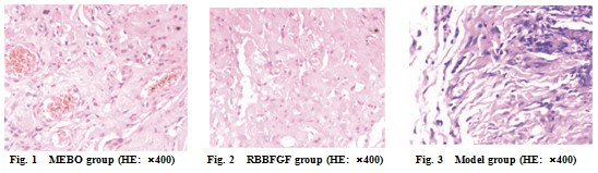
2.3. Results of optical microscope examination
On the 8th day after the administration of medication in MEBO group, granulation tissue on the wound bed was growing actively with the existence of abundant capillaries. Fibroblasts and collagenous fibers could be inspected around capillaries. The epithelial cells around the wound bed were extending and creeping in the direction of the wound bed. On the 8th day after the administration of RBBFGF in RBBFGF group, the growth of granulation tissue could be inspected. The growth of capillaries was less abundant than MEBO group, and the difference was apparent. On the 8th day after administering medication in model group, a handful of capillaries were inspected with much more local exudates and abundant inflammatory cells (see fig. 1 to fig. 3). Numbers of blood capillaries in MEBO group, RBBFGF group and model group were 12.13±2.26,9.87±1.99,7.60±1.50 respectively. The comparison of MEBO group to RBBFGF group P=0.007, P<0.01 had apparent difference with statistical significance; the comparison of MEBO group to model group, P=0.0000, P<0.01 had significant difference with statistical significance (see table 2).
Table 2 Comparison of new capillary count on the 8th day after the administration of medicaiton(x±s)
Group
n (No.)
Counts of capillary
MEBO group
15
12.13±2.26
RBBFGF group
15
9.87±1.99▲▲
Model group
15
7.60±1.50**
Notes: ▲▲Compared to MEBO group P<0.01; ★★compared to MEBO group P<0.01.
2.4. Results of electron microscope examination
Results of electron microscope examination after 8 days of interventions displayed abundant fibroblasts with large soma and regular keryon were inspected at the same location in MEBO group. Mitochondria, endoplasmic reticulum, and ribosome had no apparent expansion. The alignment of collagenous fibers with proper diameter was in order. The condition in MEBO group was slightly better than that in BRRFGF group and apparently better than model group (see fig. 4 to 6). The numbers of mitochondria in vascular endothelial cells of the wound bed in MEBO group were much more abundant than the other two groups, with integral structure, appearing round or oval. The ridge structure inspected was integral and clear. In RBBFGF group, structure of mitochondria in vascular endothelial cells was slightly worse. In model group, number and structure of mitochondria were the least in number and in worst condition with loose, expanded, ruptured and lost mitochondrial ridge from swelling. (See fig, 7-9).

3 Discussion
The repair of skin ulcerative wound is a complicated but order pathological process, consisting of hypertrophy of granulation tissue. Granulation tissue is constituted by abundant capillaries, micro-vessels and ample fibroblasts with infiltration of inflammatory cells. The formation of granulation tissue replenishing new tissues on wound, is beneficial for the anti-infection capacity, the contraction of granulation tissue, and the healing of the wound, creating the essential conditions for epithelial creeping. The granulation tissue consists of fibroblasts and new blood capillaries. The reinforcement of the proliferation capacity of capillary endothelial cells and fibroblasts can apparently promote the hypertrophy of granulation tissues. Meanwhile, the cells can excrete many functional proteins that can promote the capabilities of wound contraction and expansion [6]. Fibroblast is the key cell in the wound repairing that is known as ‘engineer, builder and manager’ of wound repairing. It can produce multiple extracellular matrix and cell factors, promote the synthesis of collagen and regulate the whole process of wound repairing. After injury, under the regulation of cell factors, fibroblasts are activated to proliferate, migrate, synthesize and secrete collagen, extracellular matrix and collagenase, which are involved in and play important roles in formation of granulation tissue and the process of contraction of wound and tissue reconstruction. Change in number of fibroblasts will have significant impact on wound healing[7].
MEBO Wound Ointment (MEBO) consists of sesame oil and beeswax as the base, then Scutellaria Baicalensis, Golden Cypress and Rhizoma Coptidis are added to clear heat, detoxicate and eliminate dampness. Angelica Sinensis and earth worms can promote the circulation and remove blood stasis. With the synergic reaction of Scutellaria Baicalensis and Angelica sinensis as the formula of Danggui Buxue Tang Decoction, it can supplement ‘qi’ and blood and promote the growth of granulation tissue. Technologically advanced equipments are used to manufacture, a unique dosage form of net-frame structure with oil enclosed by beeswax. MEBO’s net-frame dosage form can change with temperature change. At room temperature, MEBO Wound Ointment resembles semi-solid soft ointment. After being smeared on the wound bed, MEBO Wound Ointment can transform to liquid due to skin temperature. Plant oils with great lipotropic power, contained in MEBO Wound Ointment, allow liquefied ointment react with necrotic tissue. The ointment then loses its lipotropic properties and shift out of the wound bed after being mixed with exudates and liquefactions on the wound bed. With the continuous permeation of ointment from the top layer downward, the liquefied necrotic tissue and the ointment at the bottom layer are unceasingly expelled out of the wound bed. This circulation guarantees necrotic tissues are liquefied from the outer layer to inner layer and discharged without secondary damage.
This study demonstrates that MEBT/MEBO can apparently shorten the healing time of experimental SD male rats with chronic refractory wounds and have the effect of improving the healing of rats with chronic refractory wounds. As indicated in this study, MEBT/MEBO can regulate the fibroblasts on the wound bed to promote the division and proliferation of fibroblasts, promote the growth of newly grown capillaries, accelerate the formation of granulation tissues, improve the micro-circulation on the wound bed and regulate the formation of repairing matrix on the wound bed, thus enhance the capability of wound repairing and promote wound healing. This study enriched and developed MEBT/MEBO, provided the experimental evidence for the new application of MEBO Wound Ointment and the theoretical evidence for the clinical application of MEBT/MEBO.
References
[1] TANG Qian-li, Guo Lu, Wang Quan-sheng et al. The experimental study of MEBT/MEBO to rough endoplasmic reticulum of vascular endothelial cells [J]. The Chinese Journal of Burns Wounds & Surface Ulcers, 2010, 22 (4): 252-257.
[2] LU Dong, GUO Lu, TANG Qian-li et al. The therapeutic observation of MEBT/MEBO in treatment of chronic refractory ulcers [J]. The Chinese Journal of Burns Wounds & Surface Ulcers, 2010, 22 (5): 346-350.
[3] TANG Qian-li, ZHANG Li, WU Song-he et al. Endeavour to develop scientific research work and popularization of standardized MEBT/MEBO---sum-up of “Research of Popularization of Standardized MEBT/MEBO Application” [J]. The Chinese Journal of Burns Wounds & Surface Ulcers. 2008, 20 (1): 1-5.
[4] FU Xiao-bing, SUN Tong-zhu, SHENG Zhi-yong. Several animal models in the use of wound repairing study [J]. Chinese Journal of Experimental Surgery, 1999, 16 (5): 479-480.
[5] XU Rong-xiang. Clinical Handbook of Burn Regenerative Medicine and Therapy [M]. Beijing: Taihai Press, 2006: 79-80.
[6] FU Xiao-bing, TIAN Hui-min. New progress of oversea wound study [J]. Foreign Medicine Science. Osteology, 1994, 15 (3): 147-152.
[7] QIN Quan-jiang. Fibroblast function and regulation in wound healing [J]. Foreign Medicine Science. Foundamental Concerns of Traumatic Wound and Surgery, 2000: 12 (1): 33.
Received May 18, 2011