- Clinical application of MEBT/MEBO in the treatment of electric burn

-
目录
- 再生医疗技术治疗电烧伤的临床应用研究
- Clinical application of MEBT/MEBO in the treatment of electric burn
- 烧伤样本的临床特点及其表达
- MEBO在大面积烧伤晚期残余创面的临床应用
- MEBO包扎疗法治疗烧伤残余创面疗效观察
- 烧伤创面外用药物诱发局部过敏反应的临床处理
- 精神分裂症合并烧伤的临床护理
- 硫普罗宁对烧伤患者肝功能异常的治疗作用
- 湿润烧伤膏治疗手指末节皮肤软组织缺损的临床观察
- Clinical Observation of MEBO in Treating Skin Soft Tissue Injury of Distal Finger
- 人可调节封闭式负压引流技术治疗外伤创面的临床效果观察
- 美宝创疡贴治疗创伤性皮肤软组织缺损临床体会
- 美宝创疡贴治疗手术切口的疗效观察
- 湿润烧伤膏配合手术治疗合并组织缺损的手掌贯通伤
- 湿润烧伤膏配合加味骨碎补汤促进骨折愈合的临床研究
- 美宝创疡贴治疗骨折患者皮肤软组织挫擦伤
- 湿润烧伤膏在激光治疗静脉曲张术中和术后的临床应用
- 痔疮术后局部综合换药治疗的疗效分析
- 面部瘢痕的手术治疗
- 湿润烧伤膏联合美宝创疡贴在皮肤创疡治疗中的应用
- 美宝创疡贴联合湿润烧伤膏治疗慢性溃疡的临床观察
- 湿润烧伤膏联合远红外线治疗压疮的疗效观察
- MEBO外用联合口服芪黄通络汤治疗足踝部损伤性溃疡21例疗效观察
- 湿润烧伤膏治愈盲肠残端瘘1例报告
- 湿润烧伤膏治疗阑尾炎术后切口感染观察
- 湿性医疗技术与常规方法处置慢性肥厚性鼻炎激光术后创面效果比较
- MEBO处理宫颈环形电切术后创面的疗效观察
- 肝细胞移植解决α1抗胰蛋白酶缺乏
- Wild-type Hepatocytes transplantation for α1-Antitrypsin deficiency
HU Dong-cai, HU Qing-quan, LI Fan
Affilitions: Chinese Burn Association of the Integration of Traditional and Western Medicine, Beijing 100020 China (HU Dong-cai);The Graduate School of Medical College of Nanchang University, Nanchang 330006, Jiangxi Province, China (HU Qing-quan);The First People’s Hospital of Guang’an City, Guang’an 638550, Sichuan Province, China (LI Fan)
【Abstract】 Objective To introduce the clinical manifestations, debridement, wound care and management of various kinds of electrical burns. Methods Timely use MEBT/MEBO in the treatment of electrical burns on different parts of body and emphasize the attentions in the course of clinical interventions. Results MEBT/MEBO can protect the viable tissue in the occlusive layer after electrical burns to the maximum. And electrical burn injuries on all over the body targeted in this article can be regeneratively healed. Conclusion MEBT/MEBO is a safe and feasible method with reliable therapeutic effect and lower disability rate in the treatment of electrical burns.
【Key words】 Electrical Burns; Clinical Treatment; MEBT/MEBOElectrical burns can be classified into the hot burns caused by electric arc and the electrical contact burns caused by the electrical current into human body. The former is the skin burns caused by electrical spark; its characteristic is basically the same as the scalding. The latter covers the muscular, nervous, vascular, internal organ and bone injuries caused by the heat converted from electrical current that enters into human body after the direct contact with high-voltage power. The electrical resistance is different for different parts and tissue organs in human body. As a result, the injured extension is different when electrical current is passing through human body. The value of electrical resistance of each tissue is implicated with water content; the largest is bone, then adipose tissue, tendon, skin, muscle, vessel and nerve. Besides, the severity of injuries is intimately related with the intensity of electrical current and the touching duration. The clinical manifestations of arc burns are roughly the same as scalding, the damage mainly on the skin. The entry of high-voltage electrical burns appears centrically charred forming a hollow crater wound bed. The wound bed seems like the port of calabash surrounded by grayish or tan hard necrotic skin. The outer layer of wound bed appears blackish or bright red shrunk ring with slightly protruding margin. Dead muscle, vessel and nerve with large area can commonly be inspected at the center of wound bed. For severe cases, the charred dead bone could be inspected in deep tissue. The entry wound might be one or more and is bigger than the outlet wound. The sizes have no proportional relationship between both of them. Most of the outlet wound are round and dry, caused just like ‘the outburst’ from the expelling current. It appears dry and charring with loss of sensation. The middle of outlet wound is hollow or spotted. Therefore, burn surface area could not be considered as the only gauge in the severity of electrical burns. The patient suffered from high-voltage electrical burns is susceptible to the onset of disseminating injuries that means multiple outlet wounds. For upper extremities, the multiple outlet wounds could be found at armpits, elbows and wrists; for lower extremities, the popliteal space and groin are the frequent wound outlets. But, the integrity of skin tissue between outlets is maintained. The muscular death between outlet wounds caused by high-voltage electrical burns is one sort of disseminating injuries. In clinic, to perform the thorough debridement of necrotic tissue is not an easy work.
1. Early interventions
1.1. The on-the-spot first aid and wound assessment
The victim should be firstly moved from electrical power. For those with respiratory or cardiac arrest, CPR should be administered immediately. And then rapidly obtain the victim’s medical history and a series of information concerning the sort of electrical current, the locations of entry and outlet wound and etc. In addition, whether the head injuries, internal organ injuries, fracture, pneumothorax occur or not should be alert so as to make definite diagnosis via further examinations and give corresponding interventions.
1.2. Fluid replacement and anti-shock therapy
Due to the extensive muscular injuries accompanied with electrical burns and the small port of wound bed (the so-called bottle like wound with small port and wide bottom), the exudates mostly occur at the interstitial space that is disproportionate with the size of wound bed. So the infusion amount is usually underestimated. Therefore, the amount of fluid replacement could not only be calculated in accordance with total burn surface area, but increased or adjusted in accordance with specific conditions on the basis of common principle. Generally, patient should be not agitated, but has stable respiration and heart rate should be lower than 120 beats/min with strong pulsation of arteria dorsalis pedis and warm distal extremities. In particular, for those with hemoglobinuria, the fluid is intravenously administered to restore blood volume. In addition, Mannitol should be given at the same time to prevent the block of kidney tubules. And the urine output should be maintained from 200 ml/h to 300 ml/h at the first 24 hours; the alkaline medications should be given in meticulous consideration. After that, urine output should be maintained between 70 ml/h-100 ml/h. When diuresis begins and when hemoglobin disappears in urine should be recorded. If hemoglobin is persistently found in urine, it indicates the large-scale muscular necrosis. At this time, the surgical exploration should be performed on affected area timely such as the burned extremities. For those with head injuries in association with cerebral edema or persistent coma, the ice bag can be offered to decrease the temperature or 50% Glucose Solution 60 ml~100 ml can be administered via I.V. route. Meanwhile, as a result of low blood pressure after electrical shock, myoglobinuria and the direct effects from the toxin secreted by necrotic tissue and electrical current, it should be paid attention to alkalize the urine and accelerate the urine output to protect renal functions at early stage.
1.3. Early debridement
The patients with electric shock are commonly complicated with severely cardiac, cerebral and renal disorders. After the admission, the interventions including anti-shock, cardiac and renal protection should be firstly carried out. After the basic stabilization of overall conditions, the debridement should be performed during which necrotic tissue and those with deep II degree burns should be removed firstly. And then extend the wound bed from the distal to proximal to fully expose the burned deep tissue. For the ones with muscular necrosis, the anatomic continuity of nerve and tendon should be maintained as much as possible. During the debridement, the muscular tissue that appears edema and slightly pale, but has the retraction when being incised or bleeding actively should be preserved. The nerve or muscle tissue with dark redness, without contraction when being incised, apparently liquefied, infected and dead should be excised without secondary damage to adjacent viable tissue. After the debridement, the wound bed should be administered MEBO Wound Ointment at a film no more than 1 mm and performed dressing-change once per 4 to 6 hours to promote the continuous liquefaction and discharge of necrotic tissue. After the growth of granulation tissue, the autonomous skin-cluster grafting is applied to promote wound healing. Ma Yinzhen et al applied MEBT/MEBO in association with early debridement in treatment of severe electrical burn injury and obtained outstanding therapeutic effects. Compared to the other ways, the extremities were extremely protected[1].
1.4. Non-invasive fasciotomy
After high-voltage electrical burn injury, deep tissue dies with oozing of large amounts of body fluid that leads to edema beneath fascia. And then the high pressure causes the dysfunction of venous return. The increased venous pressure will aggravate and accelerate the death of muscle and other tissue. Therefore, if the edema on extremities swells apparently with progressive aggravation, non-invasive fasciotomy should urgently be performed to reduce the local pressure to avoid the fascial high-tension as a result of muscle swelling. The carpal canal should be completely incised for the burned wrist, either MEBO Wound Ointment or MEBO Gauze is smeared onto or stuffed into wound bed and then wrap up with sterile dressings. And in clinic practice, it must be noted that the extension of fascial incision should be sufficient. The tension must be released completely. It follows the principle of no bleeding during incision to attain the effect of tension-releasing.
1.5. Prevention of massive hemorrhage
The injury on deep wound causes the impairment of large vessel. Both the elevation of blood pressure after the correction of shock and the dissolution of necrotic tissue could lead to the rupture of big vessel and then massive hemorrhage. Therefore, the impaired vessels should stably be fastened during the performance of fasicotomy and the surgical kits and tourniquet should be prepared at the bedside in order to prevent urgent status after massive hemorrhage. The affected area should be observed if there occurs oozing of the blood or the aggravation of blood oozing in regular interval. When patient is extremely restless, the hemorrhage as a result of the rupture on the suture should be vigilant, especially uneasily noted in the night. If it could not be found, shock and even death in short-term period would be resulted. Hence, the patient must be observed frequently in the night. When the massive hemorrhage is found, the correct hemostasis should be provided timely.
2. Wound treatment
2.1. Electrical arc burns
The electrical arc burns mainly affect the exposed parts, generally including hands, face and/or forearms. The major reason is the near distance of hands to the electrical spark. The charred appearance of affected skin as a result of high temperature is misunderstood as deep burn injury and then carried out the surgical procedures. As a matter of fact, although the temperature of electrical arc is extremely high, most of the affected part does not locate the center of arc. And the persistent period of electrical art is quiet short, electrical arc burn injury is commonly much less to cause deep burns. For the treatment of mild electrical burn injury by electrical arc, directly apply MEBT/MEBO. Generally, the superficial layer of necrotic tissue could be automatically shed in one week with the growth of smooth and pinkish epithelium. At the later stage, there would have occurred mild hyperpigmentation. But the skin color will be restored in one month. For the treatment of severe burn injury, it should perform ‘skin-incision and tension-relieving’ technique prior to the application of MEBT/MEBO. Moreover, before topping up MEBO Wound Ointment, liquefactions attached on wound bed need to be removed till wound healing.
2.2. Low-voltage electrical burn injury
For the patient with low-voltage burn injury admitted from emergency room, shock or combined injuries should be treated actively if the patient is associated with shock or combined injuries. After the stabilization of patient’s condition, surgical procedures (such as debridement, surgical exploration) can be given to remove necrotic tissue and protect the viable tissue in the occlusive layer; if the affected limbs are found edema and hardness, it is better to perform fasciotomy to restore the blood flow and decrease the chance of amputation. For the ones admitted for a long period after injury or admitted without being given debridement as a result of overall negative conditions, the elective debridement needs to be performed. After the debridement, directly smear MEBO Wound Ointment on the superficial burns and follow the standard procedures of MEBT/MEBO. For deep wound that is unable to smear MEBO Wound Ointment, MEBO gauze could be chosen to stuff the wound bed and change the dressing every 12-24 hours to maintain the patent drainage of wound bed. With the gradual progress of liquefaction, necrotic tissue becomes less and less. Generally all necrotic tissue will be liquefied and discharged within 15-20 days. For deep II degree burns or small size III degree burns, keep using MEBT/MEBO till wound healing. For the large size III degree burns, either autonomous skin-cluster implantation or flap transplantation can be chosen to close the wound after the growth of fresh granulation tissue.
2.3. High-voltage electric shock
High-voltage electric shock often causes the impairment into deep tissue (such as vessel, nerve and muscle) due to the electric current passing through human body. The impairment extension and range in deep tissue are various and complicated. Due to secondary necrosis especially on muscle tissue caused by the progressive micro-vascular block that brings great difficulties to discriminate the boundary between burned tissue and viable tissue as well as to determine the depth of escharectomy. We can use inspection, viable tissue staining technique and methylene blue test to discriminate the viable tissue from impaired tissue. Burned muscle appears pale or dark redness with edema. No contraction will be inspected after stimulated by forceps holder, incision or direct current. Applied with tourniquet, blood can be not evacuated as a result of vascular thrombosis. Compared to normal muscle, the impaired muscle appears redness. But viable muscle appears pale and ischemia after the blood evacuation from vessel; Frozen section of viable tissue could give more accurate assessment. But its clinical use is minimal as a result of long waiting time. In addition, methylene blue test can also discriminate necrotic tissue from viable tissue: 24 to 48 hours before debridement, inject 2 ml~4 ml methylene blue beneath the eschar. During the debridement, the color of necrotic tissue will be inspected blue and will not be diluted even washed by normal saline; however, the viable tissue generally will not be stained. Due to the blood circulation, the stain agent absorbed will be discharged in urine.
For high-voltage electric shock, debridement should be given as soon as possible after the stabilization of patient’s conditions in order to remove negative effects from the toxin generated during the process of decomposition of necrotic tissue that might cause the purulent infection and be the hotbed of anaerobic bacteria. Generally, debridement should be administered within 3 to 10 days after injury. The chance of infection would be increased after 10 days that is susceptible to lead to secondary damage. After the debridement, wound should be sealed with skin-flap, muscle-flap or skin-muscle flap. For the deep wound with much necrotic tissue, the use of moist semi-exposed therapy or loose moist bandage therapy with the stuff of MEBO gauze and the perfusion of MEBO Wound Ointment is appropriate. For the severely damaged great vessel, with the liquefaction and removal of necrotic tissue by the action of MEBO gauze, it is susceptible to cause vascular rupture and massive hemorrhage. Therefore, suture kits should be prepared at bedside. Bear in mind that it is saver to perform the vascular ligation 2 cm above the normal level.3. The characteristic and management of electrical burn injury on special parts
3.1. Electrical burn injury on skull
The outer scalp covering the skull has compact and dense structure. Its characteristic is thick epithelium with large amounts of hairs deeply anchoring into the dermal layer. The blood circulation is abundant in scalp that has great power of self-healing. The external circumferential lamella of the skull is exposed and dried to death that is caused by the death of the whole scalp and the superficial layer of periosteum on the external circumferential lamella after high-voltage electric shock. But as a result of the buffering from the diploe bone between internal and external circumferential lamella, it generally will not cause the death of the entire skull. Owing to the absorbability of dead skull, it is not suggested excising them. The purpose of wound healing can be attained through the growth of new bone after the gradual absorption of dead lamella by the way of MEBT/MEBO.
In the recent years, MEBT/MEBO has been routinely using in the treatment of skull electrical burn injury. After growth of fresh granulation tissue cultivated by MEBT/MEBO fully covers the wound bed, skin-grafting is applied by which better therapeutic effect is gained. The detailed procedures as followed: drill the holes on external circumferential lamella till bleeding of diploe. The diameter of driller should be 0.2 cm~0.3 cm. The drilling interval is 0.5 cm. It is unnecessary to excise the dead skull between the drilling holes. Apply MEBT/MEBO, and then skin-grafting will be used to close the wound bed after the granulation tissue has grown abundantly on wound bed. The dead skull diploe will be absorbed. And then new bone is able to be regenerated to avoid the potential complications and sequelae as a result of skull defects; for small size electrical burn injury (within 5 cm2), directly transplant autonomous skin flap to repair the wound after the removal of dead diploe; for those with protrusion of brain parenchyma, MEBT/MEBO can be used on the surface of brain parenchyma for small size wound. After growth of abundant granulation tissue, skin-grafting can be applied. The above procedure has short-term course of treatment and less complications. Hence, the primary healing can be gained.[3] It is noted that cautious attention must be paid during the wound opening and the removal of necrotic skull. And also can the local fasical flap (or skin flap, or rib frame) be used to cover wound and then perform skin-grafting. And then steel cap is able to be used in the protection of wound bed.
3.2. Electrical burn injury on face
It is common to see the electrical burn injury on face. Due to the abundant blood circulation and loosen tissue structure on face, the onset of shock and infection will be increased with moderate edema. And the facial burn injury commonly associates with respiratory burn injury. Therefore, interventions should be timely carried out including tracheotomy and tracheal intubation, clearance of airway and maintenance of patent airway. Generally, escharectomy should not be given at early stage, but within 2-3 weeks after injury. By the action of MEBO Wound Ointment, eschar will be discharged and granulation tissue will be cultivated. At late stage, autonomous skin-cluster implantation can be used to repair the wound at late stage. The attention should be paid to prevent bleeding burst in the course of MEBT/MEBO, in particular burns on oral, lips and cheeks. The bleeding from labial arteries and facial arteries is often not obvious because of being covered by eschar. The bleeding burst occurs 7~10 days later due to the dissolution of eschar. Therefore, these arteries easily to bleed should be ligated in advance to prevent the occurrence of severe bleeding. Meanwhile, suture kits should be placed at bedside so as to carry out hemostasis timely.
3.3. Electrical burn injury on neck
The biggest danger of electrical burn injury on neck is the bleeding. For the interventions of patient with neck electrical burn injury, it is the priority to check the possibility of vascular impairment and administer the ligation on proximal vessel if necessary to prevent the unprepared massive hemorrhage. If the impairment of the common carotid artery is definite, the ruptured artery should be ligated via thoractomy. For the necrotic tissue, MEBT/MEBO should be applied as soon as possible. If MEBT/MEBO is unavailable, rotation flap can be used to cover the wound. The major complications of neck electrical burn injury are the direct vascular bleeding and indirect eschar dissolution that obstructs vascular cavity and causes secondary massive hemorrhage. In particular the bleedings on ruptured common carotid artery and internal carotid are fatal complications that should be paid high attention. Communicate with the patient and patient’s family at the early stage to gain the full understanding. At the same time, carry out necessary precautions. The rescue of patient’s life is the first priority. The restoration of better function and appearance is the second. For the III degree burn, the wound is better to be left open. Especially for the wound associated with laryngotracheal impairment, the suture cannot be given. The reason is that the inner necrotic tissue is extremely susceptible to be infected after the suture of III degree burn. Consequently, aggravate and deepen the damage or make the burned vessel rupture. After the sealing of wound, saliva of posterior pharynx enters into the wound either that increases the local physiochemistric stimulation as well as propels the contaminations on wound bed to block airway.
3.4. Electrical burn injury on chest
The patient with abroad defects of chest wall, through chest wall or thoracic opening injured by high-voltage electricity, their conditions are complicated and fluctuated with high rate of mortality. For the ones associated with open pneumothorax, sterile gauze needs to be used to stuff the defects in the first-aid. Meanwhile, closed thoracic drainage should be administered at early stage to prevent the swing of mediastium. Perform thoracotomy to evacuate the air and X-ray examination to check atelectasis. If there have inner organ damage or rib injury, the dead ribs or inner organs should be excised timely. The local skin flap, the flap of latissimus dorsi muscle or rotation flap need to be used to reconstruct the wound and administer closed thoracic drainage. Besides, for the patient with open pneumothorax, it should actively administer blood transfusion and fluid replacement and correct the disturbance of water and electrolytes. Myoglobinuria is found, both alkaline medication and diuretics should be given as early as possible to protect renal functions and prevent acute renal failure. At the same time, severe disturbance of physiological and biochemistric regulation and usage lead to the change of metabolic route, increment of metabolites, increasing of protein decomposition, decreasing of protein composition and the reduced capacity to use glucose. Therefore, it is indispensible to apply nutritional support[4]. dof water and electrolytes. sion, fluid replacement and correct the imbalance of water and electrolytes. degree burn
3.5. Electrical burn injury on abdomen
The high-voltage electrical burn injury on abdomen is usually ‘outlet’ of electric current. Therefore, it is mostly combined with electrical burn injury of extremities or other parts. According to stratification of affected area, it can be classified into simple abdominal wall burn injury, whole abdominal wall burn injury and the ones in association with inner organ burn. For simple abdominal wall burn injury, follow the standard ‘skin-incision and tension-relieving’ technique or ‘thinning’ procedure with Humby’s knife on wound bed and then apply MEBT/MEBO. At late stage, use skin-grafting (the size of skin just like stamp) on the granulation wound. Most cases will be healed. For the small size wound bed, wound can directly be sutured after reconstruction.
For the ones with whole abdominal wall burn injury or combined with inner organ hemorrhage and perforation, exploratory laparotomy should be administrated as soon as possible. If prolapsed intestinal canal is found at the accidental spot, the prolapsed intestine cannot be placed back into abdomen temporarily to prevent abdominal infection from the introduction of foreign bacteria. The vital organs in danger should be removed under the repeated discussion and sufficient preparation. For the one with peritoneal burn injury, peritoneum is supposed to be excised timely, and then the stuffs made from wide fascia, titanium alloy net, a soft plain-weave silk fabric and other material can be used to protect the inner organs in order to prevent dehiscence of abdominal wall from high abdominal pressure. And then use the local skin flap to repair the abdominal defects.
3.6. Electric burn injury on extremities
High-voltage electrical burn injury has the greatest chance to affect the extremities, generally the upper than the lower. It is mostly common to see that the upper is the entry of electric current and the lower is the outlet. The impairment of upper extremities with vital vessels and nerve affected is much more severe than that of lower extremities. For some severe cases, it could cause the death of the affected extremity. At this time, escharotomy should be performed as soon as possible to explore the impairment in association with positive interventions (promoting blood circulation). Then the amputation rate could be decreased with the recovery of partial extremity functions and reduction of deformity rate. After the stabilization of patient’s condition, skin incision and tension-relieving technique should be given as early as possible to explore and debride eschar and deep necrotic tissue. The debridement can be parted into several times and never be given after boundary of necrotic tissue is clear or muscular necrosis has been liquefied. Or it is susceptible to cause pyaemia and the progressively aggravated necrosis of the viable tissue in obstructive layer. During the debridement, the viable tissue of vital parts in obstructive layer should be retained as much as possible, including vital vessels, tendon and nerve. Use MEBO Wound Ointment to cover wound bed as early as possible. Preserve the functions of affected extremity maximally. For the case with the block of major arteries, the amputation could be administered once the victim’s life is threatened as a result of the persistent deterioration of distal vessel (appearing progressive necrosis), though fasciotomy had been given.
For the extremity wound without damaging vital vessels, nerve and tendon, apply MEBT/MEBO in combination with the debridement to excise eschar as soon as possible. The debridement can be performed without anesthesia, it is better to excise the eschar till micro-bleeding point or errhysis is inspected. Skin-grafting could also be associated to extremely protect the extremities and tissue on granulation. Take the full advantages of MEBO Wound Ointment (relax smooth muscle, release the vascular spasm and promote local blood circulation) to promote the wound healing in regeneration.
3.7. Electric burn injury on hands and wrist
Electric burn injury on hands and wrist takes up the most incident rate all over the body. And the palm is always the most severe one in hand burns. As hand often contacts and holds the electric power. The contraction of flexor group causes hand hardly to be moved from the power and results in long-term exposure under electricity. And the thick skin of palm with high electric resistance leads to the necrosis of vascular block and metacarpal bone that result in high rate of amputation. The electric burn on wrist often associates with the impairment of median nerve and ulnar nerve, but less with impaired radial nerve. Therefore, the severe deformity of wrist and fingers is accompanied in most cases.
Escharotomy should be performed at the internal and external side of wrist on wrist deep burn, including the tension-relieving of wrist and carpal canal. The arc incision should be made from the upper 1/3 inner margin of thenar to the folds of wrist, and then change the direction to the ulnar side, moving to the one-third of the distance at lower forearm in order to relieve the tension of carpal canal. If there had finger circumferential burn, the incision should extend to the digital back. In debridement, necrotic tissue should be removed in great effort to the layer of viable tissue, preserving deep flexor tendon. The burnt median nerve and ulnar nerve should be retained if no apparent liquefaction and then apply MEBT/MEBO. Electrical burn injury is susceptible to cause massive vascular impairment has certain risk to apply rotation flap.
Therefore, it is not taken as the priority[5]. Due to the thinness of the tissue around wrist, the bleeding should be noted while using MEBT/MEBO. Once it occurs, the bleeding arteriole and venule should be ligated timely. The extremity that cannot be preserved, amputation should be performed timely in one week to prevent the severe consequence from deep tissue infection. The ideal surface after amputation should be maintained in order to the future prosthesis. For the ones with healed wound, the physical therapy should be started early that is beneficial to maintain good joint position and promoter tissue repairing. The secondary transplantation of tendon and nerve on key parts can be given if necessary to preserve the certain functions of affected parts that play a key role in the improvement of life quality at rehabilitation stage.
4. Standard of wound treatment
The general principle should be followed for all electric burn injuries: Sterilize the wound with Povidine-iodine (Betadine) first; and then excise necrotic tissue (without bleeding in principle) and perform ‘skin-incision and tension-relieving’ technique with sterile scalpel; after that, apply MEBO Wound Ointment and MEBO Dressing in moist bandage therapy. The standard procedure of wound treatment is as following.
4.1. Removal of necrotic tissue without secondary damage and performance of ‘skin-incision and tension-relieving’ technique;
4.2. Any disinfectants including iodine solution, alcohol, providine iodine, hydrogen peroxide and etc should be never used on wound bed as well as normal saline. The reasons are: I. disinfectant such as providine iodine contains component of tannic acid that may cause damage on tissue cells; II. The ground substance of MEBO Wound Ointment is oil that cannot dissolve with water.
4.3. Perform the debridement without secondary damage to viable tissue as principle; no pain and no bleeding during the dressing-change. Reasons: I. avoid damaging normal tissue; II. Prevent wound infection.
4.4. Use sterile gauze to remove the wound secretions on wound bed without secondary damage;
4.5. Use MEBO Wound Ointment to smear and cover wound bed, always maintain the regenerative moist environment on wound bed;
4.6. Cover MEBO Dressing after topping up MEBO Wound Ointment and wrap up with sterile dressing till wound healing;
4.7. After the growth of granulation tissue, MEBT/MEBO can be continuously used till wound healing; surgical procedures (skin-piece transplantation, micro-skin implantation, rotation flap, pedicle flap transplantation) can also be considered if necessary. 5. Patient’s information
JMM, Male, 35 years old, from Sichuan province. The time and date of admission: 5pm, May 29th, 2010. The admitted hospital: Burn, wound and ulcer department of Guang’an People Hospital. Burn type: ten thousand volt electric burn injury on head, neck and lower extremities. Local status: The charred eschar (dead) was found on occipital area, the posterior part of neck and both foots without sensation. The admitted diagnosis: electric burn injury on head, neck and both foots with TBSA 7% (deep III degree burn).
Therapeutic procedures: MEBT/MEBO was used in the entire course of treatment. The systemic anti-infectious therapy was given after admission. By the invitation of Dr. Li (Director of burn unit), Prof. HU Dong-cai made the consultation on June 2nd,2011 and gave the orders: ‘Thinning’ procedure should be applied on wound bed to excise the necrotic tissue without secondary damage; then administer ‘skin-incision and tension-relieving’ technique. After that, change the dressing with MEBO Wound Ointment in association with MEBO Dressing, twice per day. Liquefied necrotic tissues need to be continuously removed during the treatment till wound healing. 58 days after the application of MEBT/MEBO, all wounds had been regeneratively repaired without skin-grafting. Half year follow-up showed no apparent hypertrophic scarring with good skin elasticity (See figure 1~5).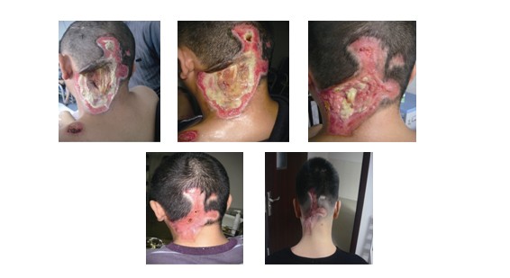
Fig.1 the 1st day after admission Fig.2 the 4th day after admission Fig.3 the 25th day after admission Fig.4 would healed at 58th day after admission Fig.5 half year follow-up
6. Conclusion
Electrical burn injury is common accidental injury that the annual incidence is about 8%~10% in statistics. It is an issue need to pay significant attention how to restore the functions of electric burn patient and decrease deformity rate. Some scholars proposed that emergent surgery should be given at the early stage after electrical burn injury[7] to maximally reduce the progressive necrosis on wound bed and preserve some nervous and tendon tissue with mild impairment that create the conditions for functional rehabilitation after wound healing. However, due to the restriction of medical level in some hospitals, many clients have to face the dilemma of amputation at last. Better therapeutic results had been gained in the treatment of electrical burn injury with MEBT/MEBO[8~ 9]. Based on effective prevention of water evaporation from wound bed, the moist environment with low-oxygen and low-tension is maintained to effectively protect wound. That is beneficial for the distension of capillaries, increment of local blood flow and prevention of the formation of micro-embolism; besides, it avoids the pressure, dryness and dehydration from bandage to make better protection and restoration of the viable tissue in obstructive layer. Consequently, preserve the extremity function to the extreme. MEBT/MEBO can effectively activate residual tissue after electrical burn injury and in situ modulate them into stem cells. By the way of continuous activation, in situ cultivation and the organic connection of stem cells with surrounding tissue, the physiological healing with good compliance is finally formed and then the promotion of tissue and skin regeneration will be gained[10]. Therefore, the application of MEBT/MEBO in treatment of electrical burn injury provides the potent security in preserving patient’s extremity, decreasing deformity rate and recovering patient’s function.Reference
[1] MA Yin-zhen, LI Jie. Experience of MEBT in Treating Electrical burn Injury [J]. Chinese Journal of Current Practical Medical, 2006, 5 (8): 68~69.
[2] DAI Yong-heng, XIE Li-ping, HE Bin, et al. The Clinical Observation of MEBT/MEBO in Association with Autonomous Skin-cluster Implantation in Treating Deep II Degree Burn on Joints [J]. The Chinese Journal of Burns Wounds & Surface Ulcers, 2009, 21 (2): 113~114.
[3] XU Rong-xiang. Complete Book of Burns [M]. Beijing: Chinese Science and Technology Press, 2008: 420.
[4] MA Wen-yuan, et al. Practical Burn Treatment [M]. Henan: Henan Science and Technology University Press, 2001: 191.
[5] XIE Wei-guo, WANG De-yun, LIU Jie-feng, et al. Use of Skin Flap in Treatment of Hand Electrical Burn Injury [J]. Chinese Journal of Burns, 2010, 26 (1): 30~33.
[6] WANG Xue, LUO Jia-xu, HANG Yan, et al. The Epidemic analysis of Patient with Electrical Burn Injury [J]. China Foreign Medical Treatment, 2010, 29 (24): 52~54.
[7] ZHANG Zhi-an, LIU Mei-chun, MAO Yuan-gui, et al. Severe Electric Burn Injury Treated by Emergent Surgery [J]. Acta Academiae Medicinae Jiangxi, 2006, 46 (4): 80~84.
[8] GUO Zhong, ZHOU Zheng-hua, Zhong G, et al. Analysis of Therapeutic Effect with MEBT/MEBO in Treatment of High-voltage Electrical Burn Injury [J]. The Chinese Journal of Burns Wounds & Surface Ulcers, 2008, 20 (4): 296~299.
[9] LI Qing, CHEN Zhong-cun, ZHAO Wei, et al. Treatment of 52 cases of electric burn [J]. The Chinese Journal of Burns Wounds & Surface Ulcers, 2004, 16 (1): 26~28.
[10] XU Rong-xiang. The Life Science Frontier-Present Status of Researhes on Stem Cell in China and Foreign Counties and Our Research Work [J]. The Chinese Journal of Burns Wounds & Surface Ulcers, 2001, 13(3): 138~159.Received Mar.15.2011